Pneumothorax
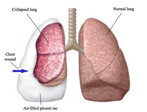
A pneumothorax is a collapsed lung, accumulation of air in the pleural space. Pneumothorax occurs when air leaks into the space between lungs and chest wall. This air pushes on the outside of a lung and makes it collapse. In most cases, only a portion of the lung collapses. A pneumothorax can be caused by a blunt or penetrating chest injury, certain medical procedures involving lungs or damage from underlying lung disease. Chest trauma carries an estimated 10-50% risk of associated pneumothorax. Sometimes, pneumothorax occurs for no obvious reason. When the lung collapses, it causes sudden chest pain and shortness of breath. A small, uncomplicated pneumothorax may quickly heal on its own. When the pneumothorax is larger, doctors usually insert a tube or needle between ribs to remove the excess air. Primary spontaneous pneumothoraces occur in young people without known respiratory illnesses. Patients with pre-existing pulmonary diseases may develop secondary spontaneous pneumothoraces. A tension pneumothorax is a medical emergency that requires immediate intervention to decompress the involved hemithorax. Patients with pneumothoraces typically complain of dyspnea and chest pain. In tension pneumothorax, patients are distressed with rapid labored respirations, cyanosis, profuse diaphoresis, and tachycardia. First-line treatment of pneumothoraces includes observation with supplemental oxygen therapy, percutaneous aspiration of the air in the pleural space, chest-tube thoracostomy, and in some cases video-assisted thoracoscopy or thoracostomy. Patients who suffer spontaneous pneumothoraces are at risk for recurrence. Pleurodesis (either by mechanical abrasion or by chemical irritation of pleural surfaces) is used to limit the likelihood of recurrence.